If you’re wondering what is on the list for minimally invasive surgery procedures, an elbow arthroscopy is definitely one of them. An elbow arthroscopy is a surgery procedure that utilises a tiny device called an arthroscopy. To understand how an elbow arthroscopy is performed, it’s important that we first understand what makes up what joints and tissues make up the elbow.
In the elbow there are two round protrusions called the epicondyles, this is where the muscles attach and the lower end of the humerus flares into. The ulna refers to the upper end of this section and there are also two protrusions known as the olecranon. The olecranon forms the point section of the elbow. These protrusions all fit into two related depression sections at the lower end of the humerus and create a hinge-like joint known as the humeroulnar. It is this joint that allows you to bend and straighten your arms.
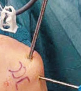
If you were to visit a Melbourne clinic specialising in elbow arthroscopy, you will find that the doctors will recommend an arthroscopy procedure to treat stiffness, arthritis, tears, and fractures in the elbow. The first step in the surgery is for your surgeon to look inside your elbow. This will be done by making small incisions and inserting thin instruments to evaluate your elbow condition. Patients won’t feel any pain as this process is done under anaesthesia. After the surgeon has inspected your elbow, they will insert thin operating tools which will be monitored using a camera so the surgeon can make the necessary repairs.
Many arthroscopy procedures, including shoulder arthroscopy, have distinct advantages. As the surgery utilises tiny holes and tiny instruments as opposed to open surgery, the patient will have a lot less scarring and soft tissue around the structures of the joint will be less likely to be disrupted. Patients can also expect a shorter stay in hospital and a faster recovery time.